
Fig. 4
- ID
- ZDB-FIG-170222-60
- Publication
- Reade et al., 2017 - TAEL: A zebrafish-optimized optogenetic gene expression system with fine spatial and temporal control
- Other Figures
- All Figure Page
- Back to All Figure Page
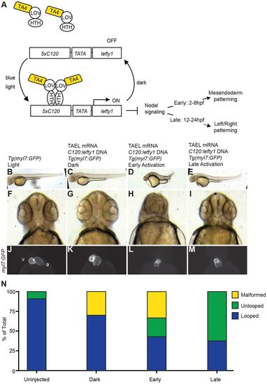
Temporal control of Nodal signaling via TAEL-induced expression of lefty1. (A) Schematic depicting experimental set-up for TAEL-induced Nodal inhibition. Embryos were injected with a transgene containing the C120 promoter driving expression of the Nodal antagonist lefty1 along with TAEL mRNA. Embryos were then globally illuminated with blue light either from 2-8 hpf (early) or 12-24 hpf (late). (B-E) Lateral views of embryos at 48 hpf show that early activation results in severely shortened embryonic axis (D) whereas late activation does not affect body length (E). (F-I) Rostral views of 48 hpf embryos show that early (H) but not late (I) activation produces cyclopia, indicative of loss of cephalic mesoderm. (J-M) Ventral views of 48 hpf embryos with hearts labeled by Tg(myl7:GFP) expression. Control embryos exhibit asymmetric heart looping (J,K). In contrast, late activation of lefty1 expression produces unlooped hearts with both chambers located at the midline (M). Early activation produced both unlooped and malformed hearts (L). A, atrium; V, ventricle. (N) Quantification of heart defects. Uninjected, n=55; dark, n=10; early, n=21; late, n=24. |