
Fig. 5
- ID
- ZDB-FIG-161229-34
- Publication
- Galant et al., 2016 - Embryonic origin and lineage hierarchies of the neural progenitor subtypes building the zebrafish adult midbrain
- Other Figures
- All Figure Page
- Back to All Figure Page
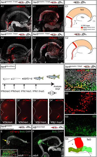
Post-embryonic her4-positive tectal RG are neurogenic during a brief window of competence linked with their position close to the TPZ. A-D. Compared distributions in the adult TeO of the progeny of her4- (A1, A2) and her5- (B1, B2) positive cells, traced from 1dpf (A1, B1) or 5dpf (A2, B2) by means of Cre-mediated recombination in her4actswith and her5actswith animals. Confocal images of horizontal sections at equivalent dorso-ventral positions, anterior left, immunostained for mCherry expression and counterstained with DAPI. Red arrows in A1, A2 point to the posterior limit of the her4-derived mCherry-positive stripe. Pink arrows in B1, B2 point to the anterior limit of the her5-derived mCherry-positive domain. Note that the position of these limits recedes towards posterior as recombination is induced at later stages, and that the her4-derived stripe is consistently anterior to the her5-derived domain. C1 and C2 are interpretative drawing (color-coded, see key) of superimposed her4actswitch and her5actswitch recombinations at 1dpf and 5dpf, respectively. D. Top panel: 9TB induction and analysis time points for the her4H2a-mCherry animals photographed in D1-D4. Bottom panels: Whole-mount dorsal views of TeOs in adult her4H2a-mCherry fish induced between 2 dpf and 1.5mpf, observed for H2a-mCherry expression under a fluorescence binocular. TeO surrounded by dotted line, anterior left, arrows to the H2a-mCherry-positive stripes. Note the receding position of these stripes with later induction time points. E. Compared antero-posterior positions in the adult TeO of an her4actswith, T(5dpf) -recombined stripe (red) and BrdU-positive cells pulsed at 5dpf (green). Yellow arrow to the identical location of the two stripes. Horizontal section observed by confocal microscopy, immunostained for mCherry and BrdU, and counterstained with DAPI. F. Antero-posterior position in the adult TeO of BrdU-positive cells pulsed at 15dpf (green). Horizontal section observed by confocal microscopy, immunostained for BrdU and counterstained with DAPI. Note the coincidence with the anterior limit of the her5actswith,T(5dpf) domain (pink arrow in panel B2). G. High magnification of a her4actswith, T(5dpf) -recombined stripe in the adult TeO, co-immunostained for mCherry and GS and counterstained with DAPI. Confocal views of a horizontal section, anterior left, yellow arrows to the mCherry-positive stripe. G4 is an interpretative drawing of G1 with identical color code. From anterior to posterior: anterior RG cells (yellow asterisk) are not overlaid with mCherry-positive neurons (black asterisk); RG at the level of the stripe (yellow asterisk, yellow arrows in G1) are overlaid with mCherry-positive neurons (red stripe); RG located posterior to the stripe are not recombined (green asterisk). Scale bars: A,B,E,F 100 µm, G 20 µm. |
Reprinted from Developmental Biology, 420(1), Galant, S., Furlan, G., Coolen, M., Dirian, L., Foucher, I., Bally-Cuif, L., Embryonic origin and lineage hierarchies of the neural progenitor subtypes building the zebrafish adult midbrain, 120-135, Copyright (2016) with permission from Elsevier. Full text @ Dev. Biol.