
Fig. S7
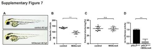
MOkcne4 testing and validation. (A) Lateral view of control- and MOkcne4 injected zebrafish embryos at 48 hpf. Note the pericardial edema and blood congestion at the inflow tract. (B, C) Quantification of heart rate (B) in control- and MOkcne4-injected embryos at 48 hpf documents a significant reduction in heart rate by KCNE4 knockdown without affecting contractility (C). (D) Luciferase reporter assay with the MOkcne4 recognizing translational start sequence from zebrafish kcne4 mRNA 5'- and in frame to the Luciferase open reading frame of the luciferase gene (pGL3kcne4). Coinjection of this construct with MOkcne4 (pGL3kcne4+MOkcne4) significantly repressed luciferase activity. Luciferase expression was normalized to renilla expression. (± sd; ***, p<0.005; three independent experiments; unpaired t-test with Welch’s correction). |