
Fig. 1
- ID
- ZDB-FIG-161129-8
- Publication
- Marro et al., 2016 - Collagen XII Contributes to Epicardial and Connective Tissues in the Zebrafish Heart during Ontogenesis and Regeneration
- Other Figures
- All Figure Page
- Back to All Figure Page
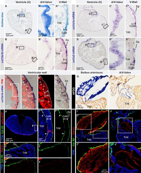
Col XII is expressed in the epicardium of the adult zebrafish heart. (A) Aniline blue staining of a ventricle transversal section visualizes collagen (blue). Framed areas encompass parts with the atrio-ventricular valve (A/V-Valve) and ventricular wall (V-Wall). The thickness of the compact myocardial layer is depicted as a bar in this and subsequent panels. Ep, epicardium; CoM, compact myocardium; TrM, trabecular myocardium. N = 5. (B-D) In -situ hybridization of ventricle sections detected by a color reaction (purple). Probe names are to the left. Framed areas encompass the parts that are enlarged in the panels to the right. N ≥ 4. (E) Superposition of a bright-field image with in -situ hybridization using col12a1b probe (purple) and fluorescent immunodetection of muscle protein Tropomyosin, TPM (red). col12a1b is expressed in the epicardium that is located externally from the myocardial border (dashed line). A few col12a1b-expressing cells are Tropomyosin-negative (arrows) and are interspersed within the compact myocardium (the thickness of the compact myocardium is indicated with a white bar). N = 4. (F) Immunofluorescence with anti-Tropomyosin (blue) to label cardiomyocytes and anti-Col XII (green) of transgenic fish wt1a(-6.8kb):GFP (red), which labels cardiac subepicardial fibroblasts (white arrows) located mainly along the junction between the outer compact myocardium (white bar) and inner trabecular myocardium. N = 4. (G, H) Aniline blue, acid Fuchsin, Orange G (AFOG) staining detects collagen (blue) in bulbus arteriosus (G, longitudinal heart section) and the leaflets of the atrioventricular valve (H, transversal heart section). N = 6. (I, J) Triple immunofluorescence staining against Col XII (green), Col Iα (red) and Tropomyosin (blue) of the structures shown in above panels. (I’) Col XII is detected on the myocardial surface. (I”) In the bulbus arteriosus, Col Iα fibers are in the interstitium, while Col XII is restricted to its surface. (J’) The atrio-ventricular connection displays Col Iα, but little Col XII in the valve leaflets. N = 6. (A’, B’, C’, D’, E’, F’) Higher magnifications of the framed areas shown in images that are labeled with the same letter without prime symbol. The same rule applies to all subsequent figures. |