
Fig. S9
- ID
- ZDB-FIG-160927-44
- Publication
- Liu et al., 2016 - Fscn1 is required for the trafficking of TGF-β family type I receptors during endoderm formation
- Other Figures
- All Figure Page
- Back to All Figure Page
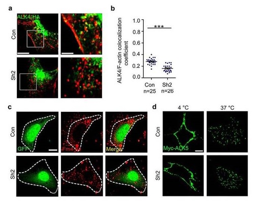
Fscn1-deletion interferes with the co-localization of ALK4 and F-actin, but does not affect the internalization of type I receptors. (a-b) NIH3T3 cells cotransfected with ALK4-HA and shRNA plasmids were fixed and costained with phalloidin-TRITC and anti-HA antibody to show the colocalization of F-actin (red) and ALK4 (green). The boxed area in the left image (Scale bar, 5 µm) is presented at a higher magnification in the corresponding right image (Scale bar, 2 µm) (a). Peason’s colocalization coefficient was quantified from the indicated cell numbers in three independent experiments and the group values are expressed as mean±SD. Student’s t test, ***P < 0.001 (b). (c) Representative fluorescence micrographs from experiments examining the uptake of FM4-64-labeled plasma membrane in NIH3T3 cells. NIH3T3 cells transfected with the indicated shRNA plasmids with a GFP marker were incubated with FM4-64 for 1 hour, fixed, and imaged using Nikon A1R+ confocal microscope system. Scale bar, 5 µm. (d) NIH3T3 cells expressing Myc-tagged ALK5 were incubated with mouse anti-Myc antibody at 4 °C for 5 h then incubated with PBS containing 10% FBS at 37 °C for 30 min. The cells were visualized by immunofluorescence with anti-Myc (green). Scale bar, 5 µm. In the colocalization analysis, NIH3T3 cells were cultured in 6-well plates. The dose of transfected plasmid DNA: Myc-ALK5, 1 µg; ALK4-GFP, 0.5 µg. |