
Fig. S2
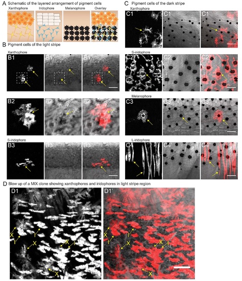
Pigment cells can be identified by their shape, color and location in the skin. (A) Schematic representation of the layered arrangement of the three pigment cell types that results in appearance of distinct light and dark stripe regions. (B) Pigment cells of the light stripe region - (B1-B1′′) xanthophores; dashed square indicates xanthophore that is enlarged in B2-B2′′, (B3-B3′′) dense and silvery s-iridophores. (C) Pigment cells of the dark stripe region - (C1-C1′′) xanthophores, (C2-C2′′) loose and blue s-iridophores, (C3-C3′′) melanophores - note the black melanin pigment, (C4-C4′′) elongated L-iridophores. In Band C: leftmost panel- fluorescence; middle panel - bright field; rightmost panel - merge. (D-D1′) Iridophores (I) and xanthophores (X) of the light stripe are distinguished by their characteristic shape. Genotype - Tg(soxlO:ERT2-Cre)/+; Tg(βactin2:loxP-STOP-loxP-DsRed-express)/+. Scale bars = 100 µm. |
Reprinted from Developmental Cell, 38(3), Singh, A.P., Dinwiddie, A., Mahalwar, P., Schach, U., Linker, C., Irion, U., Nüsslein-Volhard, C., Pigment Cell Progenitors in Zebrafish Remain Multipotent through Metamorphosis, 316-30, Copyright (2016) with permission from Elsevier. Full text @ Dev. Cell