
FIGURE
Fig. S3
- ID
- ZDB-FIG-160603-10
- Publication
- Foglia et al., 2016 - Multicolor mapping of the cardiomyocyte proliferation dynamics that construct the atrium
- Other Figures
- All Figure Page
- Back to All Figure Page
Fig. S3
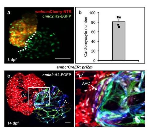
Embryonic atrial cardiomyocytes contribute to the atrial wall and atrio-ventricular canal. (a) Maximum intensity projection of a z-stack from a 3 dpf heart expressing vmhc:mCherry-NTR and cmlc2:H2-EGFP. (b) Number of atrial cardiomyocytes in 3 dpf zebrafish hearts (mean +/- SEM, n = 4, with individual data points shown). (c) Surface myocardium from a 14 dpf amhc:CreER; priZm; cmlc2:H2-EGFP heart. An expanded view of the AV canal (AVC), dense with cardiomyocyte nuclei (white), is shown. Scale bars are 50 µm. |
Expression Data
Expression Detail
Antibody Labeling
Phenotype Data
Phenotype Detail
Acknowledgments
This image is the copyrighted work of the attributed author or publisher, and
ZFIN has permission only to display this image to its users.
Additional permissions should be obtained from the applicable author or publisher of the image.
Full text @ Development