
Fig. 2
- ID
- ZDB-FIG-160524-35
- Publication
- Hackett et al., 2016 - Chemical Biology in the Embryo: In Situ Imaging of Sulfur Biochemistry in Normal and Proteoglycan-Deficient Cartilage Matrix
- Other Figures
- All Figure Page
- Back to All Figure Page
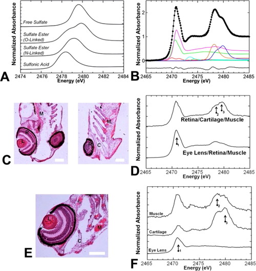
K-Edge of sulfur that is sensitive to oxidation state and chemical form. (A) Sulfur K-edge XAS spectra of model compounds representing sulfur species possible in cartilage. In particular, sulfates have different spectral features depending on their chemical form (free, N-linked ester, or O-linked-ester). (B) Least-squares fits of bulk zebrafish tissues do not detect free or N-linked sulfate, only O-linked sulfate. Color scheme: thiol, green; thio-ether, pink; disulfide, red; sulfoxide, light blue; sulfonic acid, orange; O-linked sulfate ester, dark blue. Raw data are shown as black circles, the fit is shown as a black line. The residual is shown below the spectra. (C and D) Bulk measurements performed on zebrafish tissue sections cut at different planes detect chemical differences that correlate to tissue structures present in the sample (i.e., more sulfate esters when more cartilage is present, more thio-ether when eye lens is present). (E and F) µ-XAS can be used to prove chemical speciation on a fine spatial scale (e.g., 10 µm × 10 µm region) and detect chemical differences among eye lens, cartilage, and muscle; locations from which µ-XAS (F) were measured are indicated on the histological image (E). Abbreviations for panels C and E: C, cartilage; L, eye lens; M, muscle; R, retina. Arrows numbered 1-3 in panels E and F highlight the positions of maximal absorbance due to thio-ethers, sulfonic acids, and sulfate esters, respectively, which are most abundant in the eye lens, muscle tissue, and cartilage tissue, respectively. The scale bar in panel C is 20 µm, and the scale bar in panel E is 50 µm. |