
Fig. 2
- ID
- ZDB-FIG-160512-13
- Publication
- Zhang et al., 2016 - Antiviral Drug Ganciclovir Is a Potent Inhibitor of the Proliferation of Müller Glia-Derived Progenitors During Zebrafish Retinal Regeneration
- Other Figures
- All Figure Page
- Back to All Figure Page
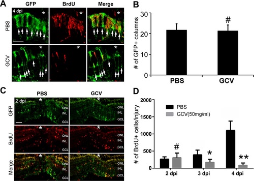
Ganciclovir had no effect on Müller glia dedifferentiation and the initial MGPC formation. (A) Confocal microscopy of retinal sections after GFP and BrdU immunofluorescence shows the presence of GFP+ Müller glia near injury site in PBS- or GCV-treated retina at 4 dpi. Tg(1016tuba1a:GFP) fish were given a pulse of BrdU 3 hours before they were killed at 4 dpi. Note that there are many fewer BrdU+ MGPCs in GCV-treated retina than in control. Arrows indicate GFP+ columns. Each GFP+ column represents an activated Müller glia and its daughter progenitor cells. (B) Quantification of the number of GFP+ columns at the injury site at 4 dpi. #No significant difference, P > 0.05, n = 4. (C) Immunofluorescence shows the initial formation of MGPCs in the INL in PBS- or GCV-treated retina at 2 dpi. Tg(1016tuba1a:GFP) fish received a pulse of BrdU 3 hours before they were killed at 2 dpi. (D) Quantification of the number of BrdU+ MGPCs per injury at 2, 3, and 4 dpi in PBS- or GCV-treated retina. Fish received a pulse of BrdU 3 hours prior to killing at 2, 3, or 4 dpi. #No significant difference, P > 0.05; *P < 0.05; **P < 0.01. n = 4 for each group. Scale bars: 50 µm. The asterisks mark the injury site (needle poke). |