
Fig. 4
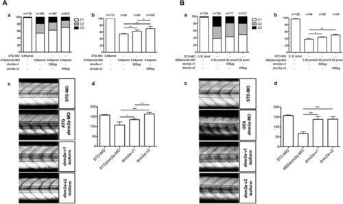
(A) Rescue with dnm2a-v1 and dnm2a-v2 after injection of ATGdnm2a-MO. (a) Appearance of embryos at 3 dpf after injection of ATGdnm2a-MO and either dnm2a-v1 or dnm2a-v2 rescue. The proportion of normal-appearing embryos (C1) is greater after rescue with dnm2a-v1 and dnm2a-v2 than after treatment with ATGdnm2a-MO alone. (b) Touch evoked response test results in normal-appearing embryos (C1) identified in experiments performed in (a). The percentage of embryos with normal touch evoked response was significantly greater in touch evoked after rescue with dnm2a-v1 and dnm2a-v2 than embryos injected with ATGdnm2a-MO alone (results obtained from 5 indipendent experiments). (c) Analysis of birefringence at 3 dpf in n = 3 somites after the end of the yolk in 6 independent replicates. Birefringence is evident in the muscle of STD-MO embryos, reduced in somites of ATGdnm2a-MO embryos and reverted to almost normal in embryos rescued with dnm2a-v1 and dnm2a-v2. (c) Graph shows quantitation of birefringence in STD-MO embryos, ATGdnm2a-MO embryos, embryos rescued with dnm2a-v1 and embryos rescued with dnm2a-v2. (B) Rescue with dnm2a-v1 and dnm2a-v2 after injection of I5E6dnm2a-MO. (a) Appearance of embryos at 3 dpf after injection of I5E6dnm2a-MO and either dnm2a-v1 or dnm2a-v2 rescue. The proportion of normal-appearing embryos (C1) is greater after rescue with dnm2a-v1 and dnm2a-v2 than after treatment with I5E6dnm2a-MO alone. (b) Touch evoked response test results in normal-appearing embryos (C1) identified in experiments performed in (a). The percentage of embryos with normal touch evoked response was significantly greater in touch evoked after rescue with dnm2a-v1 and dnm2a-v2 than embryos injected with I5E6dnm2a-MO alone (results obtained from 4 independent experiments). (c) Analysis of birefringence at 3 dpf in n = 3 somites after the end of the yolk in 6 independent replicates. Birefringence is evident in the muscle of STD-MO embryos, but significantly reduced in somites of I5E6dnm2a-MO embryos and reverted to almost normal in embryos rescued with dnm2a-v1 and dnm2a-v2. (c) Graph shows quantitation of birefringence of STD-MO embryos, I5E6dnm2a-MO embryos, embryos rescued with dnm2a-v1 and embryos rescued with dnm2a-v2. |
| Fish: | |
|---|---|
| Knockdown Reagents: | |
| Observed In: | |
| Stage: | Protruding-mouth |