
Fig. 1
- ID
- ZDB-FIG-160309-8
- Publication
- Tang et al., 2016 - Imaging tumour cell heterogeneity following cell transplantation into optically clear immune-deficient zebrafish
- Other Figures
- All Figure Page
- Back to All Figure Page
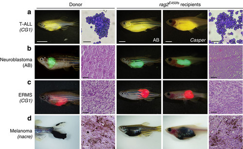
Zebrafish cancers engraft into homozygous rag2E450fs (casper) animals. Donor animals shown in the left two panels while transplant recipients are to the right. (a) ZsYellow-labelled Myc-driven T-ALL from the syngeneic CG1 background, (b) EGFP-labelled neuroblastoma from AB background, (c) mCherry-labelled kRASG12D-driven ERMS from CG1 background, and (d) BRAFV600E-induced melanoma arising in tp53-/-nacre background. Tumour cells were transplanted intra-peritoneally (a,b,d) or intra-muscularly (c) into both rag2E450fs (AB) and rag2E450fs (casper)-recipient fish. Merged brightfield and fluorescent images are shown at 30 d.p.t. Cytospins of leukaemia cells are shown in a, whereas haematoxylin and eosin (H&E)-stained sections are shown in b-d. Scale bars equal 5 mm in whole animals images, 20 µm for cytospins shown in a, and 50 µm for histology sections shown in b-d. |
| Fish: | |
|---|---|
| Condition: | |
| Observed In: | |
| Stage: | Adult |