
Fig. 4
- ID
- ZDB-FIG-160218-11
- Publication
- Taku et al., 2016 - Attractant and repellent cues cooperate in guiding a subset of olfactory sensory axons to a well-defined protoglomerular target
- Other Figures
- All Figure Page
- Back to All Figure Page
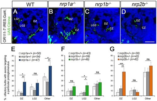
Loss of nrp1a phenocopies sema3d mutants. (A-D) Single optical sections through 72hpf or111-7 transgenic larvae (frontal view). Axons are in green. Dorsal is up and medial is to the right. Propidium iodide (blue) labels cell bodies revealing protoglomeruli as cell-free regions. WT, wild type. Scale bar: 25µm. (E-G) Percentage of OBs displaying a projection to a particular protoglomerulus or all non-protoglomerular regions (other) is shown. Statistical significance was estimated using two-tailed Fisher′s exact tests (P<0.05*, P<0.01**, P<0.001***; ns, not significant). Homozygous mutants are compared with wild-type and heterozygous siblings. Error bars represent s.e. of the sample proportion. (B,E) A subset of or111-7 transgene-labeled axons misproject to the DZ and to non-protoglomerular areas in nrp1a mutants. (C,F) nrp1b mutants do not have increased mistargeting errors to the DZ but do have increased misprojections to LG2. (D,G) A subset of or111-7 transgene-labeled axons also inappropriately targets the DZ in nrp2b mutants. CZ, central zone; DZ, dorsal zone; LG1, lateral protoglomerulus 1; LG2, lateral protoglomerulus 2. |
| Fish: | |
|---|---|
| Observed In: | |
| Stage: | Protruding-mouth |