
FIGURE
Fig. S1
- ID
- ZDB-FIG-151228-3
- Publication
- Govindan et al., 2015 - Dynamic remodeling of the extra cellular matrix during zebrafish fin regeneration
- Other Figures
- All Figure Page
- Back to All Figure Page
Fig. S1
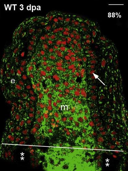
HA is upregulated in the stump of the regenerating fin. Wild type (WT), 3 dpa, longitudinal fin sections were treated biotin-HA binding protein and detected using the streptavidin conjugated with Alexa Fluor– 488 (green). Propidium iodide (nuclei) is used as the counter stain (red). White line represents the amputation plane and the asterix (**) marks the bone. The percentage of sections showing similar expression pattern is denoted in each panel (n = 40–65 sections). Arrow identifies the basal layer of epithelium (BLE); m, mesenchyme; e, epidermis; dpa, days post amputation. Scale bar is 20 µm. |
Expression Data
Expression Detail
Antibody Labeling
Phenotype Data
Phenotype Detail
Acknowledgments
This image is the copyrighted work of the attributed author or publisher, and
ZFIN has permission only to display this image to its users.
Additional permissions should be obtained from the applicable author or publisher of the image.
Reprinted from Gene expression patterns : GEP, 19(1-2), Govindan, J., Iovine, M.K., Dynamic remodeling of the extra cellular matrix during zebrafish fin regeneration, 21-9, Copyright (2015) with permission from Elsevier. Full text @ Gene Expr. Patterns