
FIGURE
Fig. 5
- ID
- ZDB-FIG-151228-15
- Publication
- Mendieta-Serrano et al., 2015 - Spatial and temporal expression of zebrafish glutathione peroxidase 4 a and b genes during early embryo development
- Other Figures
- All Figure Page
- Back to All Figure Page
Fig. 5
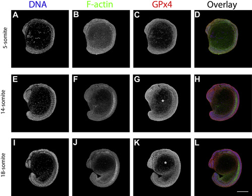
GPx4 immunofluorescence localization patterns in zebrafish embryos at different somite stages as detected by laser confocal microscopy. 5-somite stage embryos (A, B, C and D). 14-somite stage embryos (E, F, G and H). 18-somite stage embryos embryos (I, J, K and L). Hoechst-stained embryos (A, E and I). F-actin, phalloidin Alexa 488-stained embryos (B, F and J). GPx4 immunolocalization (C, G and K). Left side views are shown. *, yolk cell which start to show GPx4a signal. Scale bar 250 µm. |
Expression Data
| Antibody: | |
|---|---|
| Fish: | |
| Anatomical Terms: | |
| Stage Range: | 5-9 somites to 14-19 somites |
Expression Detail
Antibody Labeling
Phenotype Data
Phenotype Detail
Acknowledgments
This image is the copyrighted work of the attributed author or publisher, and
ZFIN has permission only to display this image to its users.
Additional permissions should be obtained from the applicable author or publisher of the image.
Reprinted from Gene expression patterns : GEP, 19(1-2), Mendieta-Serrano, M.A., Schnabel-Peraza, D., Lomelí, H., Salas-Vidal, E., Spatial and temporal expression of zebrafish glutathione peroxidase 4 a and b genes during early embryo development, 98-107, Copyright (2015) with permission from Elsevier. Full text @ Gene Expr. Patterns