
Fig. 3
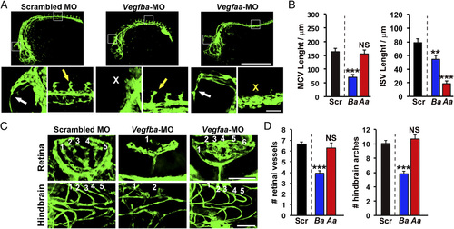
Distinct vascular defective phenotypes in VEGF-B and VEGF-A–deficient zebrafish embryos. (A) Confocal micrographs of 24-hpf fli1a:EGFP embryos injected with 0.6 pmol of Scrambled, Vegfba, or Vegfaa morpholinos. White arrows indicate the MCV, and the white X indicates the position where the MCV is missing. Yellow arrows indicate the ISVs, and the yellow X indicates the position where ISVs are missing. (Scale bar, 500 µm; bar in the amplified picture, 50 µm.) (B) Quantifications of MCV or ISV lengths in 24-hpf embryos injected with 0.6 pmol of Scrambled (Ctrl), Vegfba (Ba), or Vegfaa (Aa) morpholinos (n = 8–24 embryos/group). NS, nonsignificant. **P < 0.01; ***P < 0.001. (C) Confocal micrographs of the retina or hindbrain regions in 72-hpf fli1a:EGFP embryos injected with 0.6 pmol of Scrambled, Vegfba, or Vegfaa morpholinos. Sequential numbers indicate the number of retinal vessels or the number of hindbrain arches. (Scale bar, 50 µm.) (D) Quantification of retinal vessel and hindbrain arch numbers in 72-hpf embryos injected with 0.6 pmol of Scrambled (Ctrl), Vegfba (Ba), or Vegfaa (Aa) morpholinos (n = 7–24 embryos/group). NS, nonsignificant; ***P < 0.001. |
| Fish: | |
|---|---|
| Knockdown Reagents: | |
| Observed In: | |
| Stage: | Prim-5 |