
Fig. 3
- ID
- ZDB-FIG-151123-9
- Publication
- Li et al., 2015 - Identification and expression of Lypc, a novel dark-inducible member of Ly6 superfamily in zebrafish Danio rerio
- Other Figures
- All Figure Page
- Back to All Figure Page
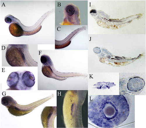
The expression pattern of lypc in early development: (A): Long-pec embryo (48 hpf), pigment cells reach terminal differentiation along dorsal strip (arrows) (B): pigment cells in the retina of embryo (48 hpf) (C): pigment cells in tail (48 hpf). (D–F): By 72 h, lypc strongly expresses on ventral side of yolk sac. Lypc positive cells have organized along the dorsal, ventral and yolk stripes. Red arrow: dorsal stripe, ventral yolk stripes (G–H). Lateral view of larva of 96 hpf. The expression pattern of lypc was mainly restricted to the ventral yolk stripes. (I–J) The longitudinal section embryos at 72 hpf after in situ hybridization (K) is showing the traverse section of embryo at 72 hpf. Strong signal in epidermal cells in the trunk and tail reveals that lypc expresses in pigment cells. (L) The retina of zebrafish embryo at 72 hpf. |
| Gene: | |
|---|---|
| Fish: | |
| Anatomical Terms: | |
| Stage Range: | Long-pec to Day 4 |
Reprinted from Gene, 574(1), Li, L., Ji, D., Teng, L., Zhang, S., Li, H., Identification and expression of Lypc, a novel dark-inducible member of Ly6 superfamily in zebrafish Danio rerio, 69-75, Copyright (2015) with permission from Elsevier. Full text @ Gene