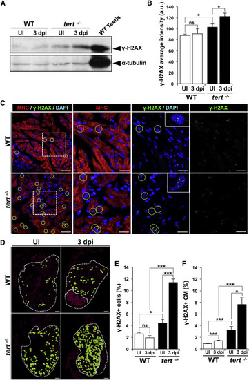
Fig. 6
|
DNA Damage Increases Strongly after Ventricular Cryoinjury in the Absence of Telomerase (A) Representative western blot of γ-H2AX expression in WT and tert-/- hearts without injury and in hearts at 3 dpi. (B) Quantification of western blot signal intensities (n = 9 hearts/condition). Data are means ± SEM. p < 0.05 (Mann-Whitney test). (C) Representative staining of γ-H2AX foci (green) in cardiac cells in uninjured and 3 dpi WT and tert-/- hearts. Cardiomyocytes are immunostained with anti-MHC (red), and nuclei are counterstained with DAPI (blue). Examples of γ-H2AX+ cardiomyocytes are outlined with green circles. Boxed areas are shown at a higher magnification. Scale bars, 20 µm. (D) Distribution of γ-H2AX-positive cardiomyocytes (green circles) in uninjured and 3 dpi WT and tert-/- hearts. The nuclear area is shown in magenta. The ventricle and injured area are outlined by dotted lines. Scale bars, 100 µm. (E and F) Percentages of (E) γ-H2AX-positive cardiac cells and (F) γ-H2AX-positive cardiomyocytes in uninjured and 3 dpi WT and tert-/- hearts. Data are means ± SEM of cells counted on a minimum of 3 sections/heart in four hearts. p < 0.05, p < 0.01, p < 0.001 (Student’s t test). See also Figure S7. |

