
Fig. 3
- ID
- ZDB-FIG-151026-2
- Publication
- Mitchell et al., 2015 - Retinoic Acid Signaling Regulates Differential Expression of the Tandemly-Duplicated Long Wavelength-Sensitive Cone Opsin Genes in Zebrafish
- Other Figures
- All Figure Page
- Back to All Figure Page
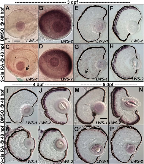
Changes in spatiotemporal patterns of expression of LWS1 and LWS2 in response to 9-cis RA treatment. A-H. Whole-mounted (A-D) and sectioned (E-H) embryo eyes obtained from embryos treated with DMSO (A,B,E,F) or 0.3 µM 9-cis RA (C,D,G,H) from 48 hpf to 75 hpf, and then hybridized with LWS1 (A,E,C,G) or LWS2 (B,G,D,H) cRNA. Arrows in C and G indicate LWS1-expressing cones in ventral retina; n, nasal; v, ventral. I-L. Sectioned embryo eyes obtained from embryos treated with DMSO (I,J) or 0.3 µM 9-cis RA (K,L) from 48 hpf to 4 dpf, and then hybridized with LWS1 (I,K) or LWS2 (J,L) cRNA. M-P. Sectioned embryo eyes obtained from embryos treated with DMSO (M,N) or 0.3 µM 9-cis RA (O,P) from 48 hpf to 4 dpf, and then hybridized with LWS1 (M,O) or LWS2 (N,P) cRNA. Scale bar in A (applies to A-H) = 50 um. Scale bar in I (applies to I-L) = 50 µm. Scale bar in M (applies to M-P) = 100 µm. |