
Fig. 3
- ID
- ZDB-FIG-150722-15
- Publication
- Lacoste et al., 2015 - A Convergent and Essential Interneuron Pathway for Mauthner-Cell-Mediated Escapes
- Other Figures
- All Figure Page
- Back to All Figure Page
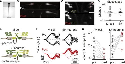
Spiral Fiber Neurons Are Necessary for Lateralized M-Cell-Mediated Escapes (A) Tail-free larvae are presented with a non-directional tap stimulus as in Figure 2. (B) Projection of two-photon image stack showing M-cells before (top) and 24 hr after (bottom) ablation of the M-cell on the left in Et(fos:Gal4-VP16)s1181t; Tg(UAS-E1b:Kaede). (C) Projection of two-photon image stack showing spiral fiber neurons before (top) and 24 hr after (bottom) ablation of spiral fiber neuron somata located on the right in Tg(–6.7FRhcrtR:gal4VP16); Tg(UAS-E1b:Kaede). The axon cap (green oval) contralateral to the targeted spiral fiber neurons is no longer apparent 24 hr after ablations. (D) Normalized change in short-latency (SL) escape probability as a function of all trials (post – pre). Gray circles, individual fish; black line, median. Left: M-cell ablation (n = 11). Right: spiral fiber neuron ablations (n = 17). The probability change is not significantly different from 0 in either condition (p = 0.67 and p = 0.98, respectively; Wilcoxon signed-rank test). (E) Model showing that when M-cells or spiral fiber neurons are ablated unilaterally, escapes in response to taps become strongly biased toward one direction: ipsilateral to the ablated M-cell or contralateral to the ablated spiral fiber neurons. (F) Example tail traces for a fish before (top plots; black) and after (bottom plots; red) ablation of the left M-cell (left plots) and a fish before and after ablations of spiral fiber neuron somata on the right (right plots). The directionality of the initial tail bend is expressed as ipsilateral or contralateral with respect to the ablated soma(ta). Traces begin at the time of tap delivery. (G) Probability of contralateral SL escapes as a function of all SL escapes of either direction. Left: M-cell ablation. Right: spiral fiber neuron ablation. Escapes shift toward the ipsilateral side for M-cell ablation and to the contralateral side for spiral fiber neuron ablations. The laterality bias after M-cell or spiral fiber neuron ablation was not statistically distinguishable (p = 0.45, Wilcoxon rank-sum test). Scale bars: 20 µm. Pictures are oriented rostral up. SF, spiral fiber; LS, short latency; contra, contralateral; ipsi, ipsilateral. |