
Fig. 2
- ID
- ZDB-FIG-150603-4
- Publication
- Kaufman et al., 2015 - Development and origins of Zebrafish ocular vasculature
- Other Figures
- All Figure Page
- Back to All Figure Page
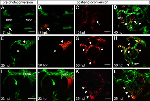
Origins of superficial and hyaloid vessels. (A,E,I) Pre-photoconversion and (B-D, F-H, J-L) post-photoconversion confocal z-stack projections of kdrl:Kaede embryos. Cells that were not photoconverteed are green whereas photoconverted cells are red. Age of embryos when imaged is depicted in each panel. (A-D) Photoconversion of left ROC. (A,B) Arrows point at the left ROC. (C,D) Single channel image showing only photoconverted endothelial cells (C), and a merge of channels showing photoconverted and non-photoconverted cells (D). Arrows and arrowheads point at central hyaloid vessels with photoconverted cells and more peripheral hyaloid vessels without photoconverted cells, respectively. (E-H) Photoconversion of PMBC. (E,F) Asterisks mark the center of the eye. (G,H) Single channel image showing only photoconverted endothelial cells (G), and a merge of channels showing photoconverted and non-photoconverted cells (H). Arrowheads point at superficial vessels and arrow in H points at hyaloid vessels. (I-L) Photoconversion of ventral PMBC. (I,J) Only the ventral region of the PMBC was photoconverted (red in J). (K,L) Single channel image showing only photoconverted endothelial cells (K), and a merge of channels showing photoconverted and non-photoconverted cells (L). Arrowheads point at the VRV and peripheral hyaloid vessels. (A,B) are dorsal views, all other panels are lateral views, anterior to the left. CrDI, cranial division of internal carotid artery; DRV, dorsal retinal vessel; MOC, midbrain organizing center; NRV, nasal radial vessel; PMBC, Primordial midbrain channel; ROC, rostral organizing center; VRV, ventral radial vessel. Scale bars are 50 µm. |