
Fig. 4
- ID
- ZDB-FIG-150512-9
- Publication
- Lee et al., 2015 - IFT46 plays an essential role in cilia development
- Other Figures
- All Figure Page
- Back to All Figure Page
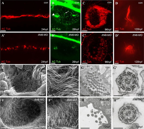
Ciliary defects in zebrafish ift46 morphants. Immunofluorescence with antibody against acetylated α-tubulin showing cilia in the pronephric ducts, (scale bar, 10 µm) (A–A′), otic vesicle, (scale bar, 10 µm) (B–B′), olfactory placode, (scale bar, 10 µm) (C–C′) and a lateral line hair cell, (scale bar, 5 µm) (D–D′). The ift46 morphants exhibit shortened or fewer cilia compared with the control fish in all of these organs. Arrow head: tether cilia. Arrow: short cilia. (E–F′) SEM images showing the cilia in the olfactory placode of control fish (E, E′) and ift46 morphants (F, F′) at 5 dpf. Scale bar, 40 µm (E) and (F), 4 µm (E′) and (F′) (G–H′) TEM images of cilia in zebrafish pronephric ducts at 72 hpf. Ultrastructure of normal pronephric cilia shows 9+2 microtubule architecture at 72 hpf which is intact in ift46 morphants. (G′, H′) Representative views of individual cilium at higher magnification. Scale bar, 5 µm (G) and (H), 100 µm (G′) and (H′). |
| Fish: | |
|---|---|
| Knockdown Reagent: | |
| Observed In: | |
| Stage Range: | Prim-5 to Day 5 |
Reprinted from Developmental Biology, 400(2), Lee, M.S., Hwang, K.S., Oh, H.W., Ji-Ae, K., Kim, H.T., Cho, H.S., Lee, J.J., Yeong Ko, J., Choi, J.H., Jeong, Y.M., You, K.H., Kim, J., Park, D.S., Nam, K.H., Aizawa, S., Kiyonari, H., Shioi, G., Park, J.H., Zhou, W., Kim, N.S., Kim, C.H., IFT46 plays an essential role in cilia development, 248-57, Copyright (2015) with permission from Elsevier. Full text @ Dev. Biol.