
Fig. 3
- ID
- ZDB-FIG-150506-27
- Publication
- Dirian et al., 2014 - Spatial Regionalization and Heterochrony in the Formation of Adult Pallial Neural Stem Cells
- Other Figures
- All Figure Page
- Back to All Figure Page
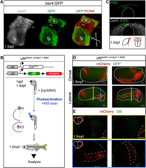
Lateral aNSCs-Fated Embryonic Progenitors Are Located in the Posterior Telencephalic Roof Plate (A) Three-dimensional view of the prosencephalon at 1.5 dpf in a her4:GFP embryo immunostained as indicated. Dashed line represents the position of the epiphysis (e). Posterior part of the telencephalic roof plate. Te, telencephalon. (B) Experimental design to fate map embryonic progenitors located at the posterior telencephalic roof plate: caged-cyclofen (cyclofen) was locally photoactivated in ubi:creErt2;ubi:switch embryos at 1–1.5 dpf using a 405 nm laser beam (blue box represents the laser-activated area). Recombined animals (ubiswitch,uncag(1-1.5 dpf)) were analyzed at 1.5 mpf. (C) Cross-section focusing on the posterior telencephalic roof plate of an embryo injected with caged-FITC and analyzed immediately after uncaging. The uncaged area (asterisks) is limited to the roof plate. (D) Dorsal (top) and lateral (bottom) whole-mount views of ubiswitch,uncag(1-1.5 dpf) telencephali. Dotted lines surround the pallium. (E) Cross-sections at the medial level (top: yellow section plane in D) and at the posterior level (bottom: blue section plane in D) of ubiswitch,uncag(1-1.5 dpf) lateral telencephali stained as indicated. See also Figure S3. |
Reprinted from Developmental Cell, 30(2), Dirian, L., Galant, S., Coolen, M., Chen, W., Bedu, S., Houart, C., Bally-Cuif, L., Foucher, I., Spatial Regionalization and Heterochrony in the Formation of Adult Pallial Neural Stem Cells, 123-36, Copyright (2014) with permission from Elsevier. Full text @ Dev. Cell