
Fig. 1
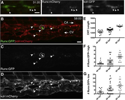
A Transgenic Zebrafish Line that Specifically Marks HSPCs (A) Single frame of time-lapse (hr post fertilization:min) showing Runx:mCherry+ HSPCs (arrowheads, red nuclei) budding from kdrl:GFP+ hemogenic endothelium of DA (green). See Movie S1. (B–D) CHT is colonized by Runx:GFP+ HSPCs (green) that are closely associated with kdrl:mCherry+ ECs (red). Caudal artery (CA) is dorsal to CHT, caudal vein (CV) is ventral, and circulation runs posterior (right arrow) and anterior (left arrow), respectively. Cluster of three Runx:GFP+ high cells outlined with dashed line and four Runx:GFP+ low cells indicated with arrowheads. 58 hpf embryo. (E–G) Runx:GFP+ high and low cells quantified, with CHT length as indicator of stage (anterior from cloaca to posterior limit of CA). Note: confocal images are 3D rendered depth or max projections of 20–30 µm z stacks. Scale bars, (A) 15 µm; (B) 25 µm. Error bars show mean ± SEM. See also Figure S1. |
Reprinted from Cell, 160, Tamplin, O.J., Durand, E.M., Carr, L.A., Childs, S.J., Hagedorn, E.J., Li, P., Yzaguirre, A.D., Speck, N.A., Zon, L.I., Hematopoietic Stem Cell Arrival Triggers Dynamic Remodeling of the Perivascular Niche, 241-252, Copyright (2015) with permission from Elsevier. Full text @ Cell