
Fig. 5
- ID
- ZDB-FIG-150325-52
- Publication
- González-Rosa et al., 2014 - Use of Echocardiography Reveals Reestablishment of Ventricular Pumping Efficiency and Partial Ventricular Wall Motion Recovery upon Ventricular Cryoinjury in the Zebrafish
- Other Figures
- All Figure Page
- Back to All Figure Page
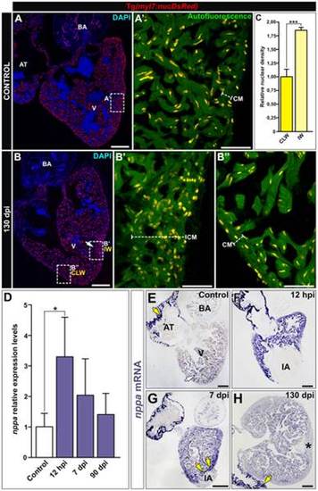
Cryoinjury induces local, long-term alterations in myocardial organization. (A,B) Immunohistochemistry on sagittal sections of control (A,A′) and cryoinjured (B-B′′) hearts at 130 dpi from the Tg(myl7:nucDsRed) line. A′-B′′ are zoomed images of boxed areas in A and B, additionally showing autofluorescence to reveal tissue organization. (A-A′) In control hearts, one or two cells constitute the thickness of the compact myocardium (CM). (B-B′′) At 130 dpi, the injured wall (IW) shows an abnormal increase in the number and distribution of cardiomyocytes compared with the contralateral wall (CLW). (C) Quantification of the nuclear density relative to the compact tissue reveals an increase in cardiomyocyte density in the IW compared to the CLW. Graph represents mean values and SD (*** p = 0.006, two tailed Student′s t-test; 100–150 cells counted per section, 3 sections per heart, n = 3 animals analyzed). (D) qPCR from ventricular RNA samples reveal induction of the natriuretic peptide encoding gene nppa upon cryoinjury. Graph represents mean values and SD, n = 4-5 replicates, Expressions levels were normalized to that of ef1α and rps11 and further normalized to that of the uninjured sample. (* p<0.05; one-way ANOVA followed by Tukey′s honest significant difference test). (E-H) Sections of cryoinjured hearts at the indicated times post-injury hybridized with a riboprobe for nppa mRNA. Yellow arrows mark areas of strong nppa expression. (E) In control hearts, nppa is highly expressed in the atrium (yellow arrow) and at lower levels in the trabecular myocardium (white arrow). (F-G) Shortly after injury, nppa is strongly upregulated in the ventricular myocardium. (H) At 90 dpi, the levels of nppa expression are similar to those detected in control hearts. Observe the increase in thickness of the compact layer of the injured wall (asterisk) revealed by no expression of nppa. AT, atrium; BA, bulbus arteriosus, CLW, contralateral wall; CM, compact myocardium; hpi, hours postinjury; dpi, days postinjury; IA, injured area; IW, injured wall; V, ventricle. Bars, 200 µm (full views), 50µm (magnifications). |