
Fig. 3
- ID
- ZDB-FIG-150120-3
- Publication
- Smith et al., 2014 - Contact-Mediated Inhibition Between Oligodendrocyte Progenitor Cells and Motor Exit Point Glia Establishes the Spinal Cord Transition Zone
- Other Figures
- All Figure Page
- Back to All Figure Page
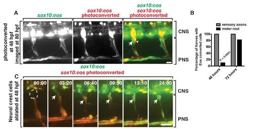
Motor root-associated glia are distinct from neural crest-derived glia. (A) In a Tg(sox10:eos) embryo exposed to UV light at 48 hpf and imaged at 80 hpf, photoconverted cells (red, arrowhead) are only observed along sensory axons while unconverted cells (green, arrow) populate the motor root. CNS and PNS portions are denoted in brackets. (B) Quantification of photoconversion experiments shows CNS-derived, unconverted sox10+ cells develop between 48 and 72 hpf, whereas converted sox10+ cells that generate the DRG are present before 48 hpf (30 nerves scored). (C) Frames captured from a 24-h time-lapse video beginning at 48 hpf in a Tg(sox10:eos) embryo that was exposed to UV light at 48 hpf and DRG ablation immediately after. Numbers in upper right corners denote stage of development. At 51.3 hpf (03:20), a CNS-derived sox10+ cell (green, arrow) associated with motor axons and generated motor root glial cells even in the presence of dying DRG cells (red). All images are lateral views of the motor and sensory root with dorsal to the top and anterior to the left. CNS and PNS portions are denoted in brackets. Scale bars, 25 µm. |
| Gene: | |
|---|---|
| Fish: | |
| Condition: | |
| Anatomical Term: | |
| Stage: | Protruding-mouth |