
Fig. 5
- ID
- ZDB-FIG-150120-23
- Publication
- Sheets et al., 2014 - Characterization of Ribeye Subunits in Zebrafish Hair Cells Reveals That Exogenous Ribeye B-Domain and CtBP1 Localize to the Basal Ends of Synaptic Ribbons
- Other Figures
- All Figure Page
- Back to All Figure Page
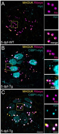
CtBP1 localizes to synaptic ribbons, but does not disrupt endogenous Ribeye. Representative images of immunolabel in posterior lateral line NM3 hair cells of 5 dpf larvae. Scale bars: 3 μm (main panels), 1 μm (insets). (A) Ribeye b antibody labeling of synaptic ribbons (magenta) and MAGUK antibody labeling of postsynaptic densities (yellow) in a WT sibling larva. Anti-myc (cyan) immunolabel was performed as a negative control. (B–C) CtBP1-myc (cyan), Ribeye b (magenta), and MAGUK (yellow) immunolabel in two representative transgenic larvae. (B) CtBP1-myc (cyan) is strongly localized to the nucleus with weak synaptic localization. The red arrow indicates a synaptic ribbon containing CtBP1-myc. Note that Ribeye immunolabel intensity in all four synaptic ribbons appears comparable. (C) CtBP1-myc (cyan) is weakly localized to the nucleus with strong synaptic localization. Presynaptic Ribeye immunolabel intensity is not reduced compared to WT (A). |