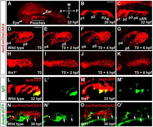
Requirement of Tbx1 for pouch morphogenesis. (A) A zoomed out view of a 32hpf her5:mCherryCAAX (red) embryo shows the position of pouches relative to the developing eye and ear. (B,C) The first two pouches (p1, p2) form by 20hpf, with the remaining pouches (p3-p6) developing at one per 4h. (D-K) Still images from time-lapse confocal recordings of wild-type and tbx1 mutant pouch development at the times indicated (bottom right of the images) (see supplementary material Movie 1A,B). her5:mCherryCAAX labels endodermal cell membranes. Recordings were started at 26hpf (T0), a stage when the fourth and fifth pouches (p4 and p5) are beginning to develop in the wild-type example. Compared with the typical pouch outpocketing behavior seen in all three wild-type siblings, no endodermal outpocketing was observed in three out of three tbx1 mutants (H-K). (L,M) Fluorescent in situ hybridization for fgf3 (green) and GFP immunohistochemistry to label the her5:GFP-positive pouch endoderm (red). fgf3 was expressed in a segmental pattern in all 39 wild-type siblings and all 14 tbx1 mutants. (N,O) Confocal sections of pre-pouch endoderm in dusp6:dGFP; her5:mCherryCAAX transgenic embryos. In wild type, dusp6:dGFP (green) was expressed segmentally in already formed pouches (arrowhead) and in clusters of presumptive pouch endoderm before outpocketing (arrows); strong expression was also seen in adjacent mesoderm (asterisks) (n=18). In tbx1 mutants, dusp6:dGFP was expressed at lower levels and in fewer cells, yet a segmental pattern in the endoderm was still detected (n=7). Note that we increased the relative gain of the green channel in tbx1 mutants to reveal the weak segmental dusp6:dGFP expression. See supplementary material Fig. S1 for the unaltered images. L′-O′ show the green channel alone. Scale bars: 40μm (B,C,L,M); 20μm (D-K,N,O).
|
