
Fig. 1
- ID
- ZDB-FIG-141125-8
- Publication
- Hartwig et al., 2014 - Temporal control over the initiation of cell motility by a regulator of G-protein signaling
- Other Figures
- All Figure Page
- Back to All Figure Page
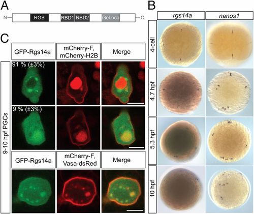
Zebrafish regulator of G-protein signaling 14a (rgs14a) RNA expression and Rgs14a protein localization. (A) A schematic representation of the Rgs14a protein structure showing the N-terminal RGS domain, the two Raf-like Ras-binding domains (RBD), and the C-terminal GoLoco domain. (B) Whole-mount in situ RNA hybridization using rgs14a (Left) and nanos (Right) antisense RNA probes at the indicated embryonic stages; 5.3- and 10-hpf embryos were overstained in the case of rgs14a in situ hybridization to detect the weak expression in the PGCs. (C) GFP-Rgs14a fusion protein expressed in PGCs is localized to the cytoplasm and the plasma membrane, with enrichment in perinuclear granules (in 91% of the 46 PGCs analyzed; Top) or in the nucleus (in 9% of the 46 PGCs analyzed; Middle). The perinuclear granules where the GFP-Rgs14a protein is localized harbor Vasa-DsRed protein (Bottom), defining them as germ cell granules. (Scale bars, 10 μm.) |
| Genes: | |
|---|---|
| Fish: | |
| Anatomical Terms: | |
| Stage Range: | 4-cell to Bud |