
Fig. S5
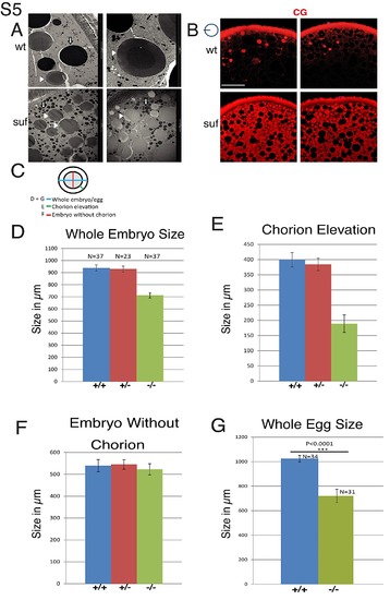
Suf/Spastizin controls cortical granule maturation. (A) Electron micrographs showing yolk globules and mature cortical granules in wt oocytes (top row). Note the darker dense core in the center of mature cortical granules. Suf/Spastizin mutant oocytes (bottom row) show immature cortical granules without dense core. Image frame: 2 μm (top, right panel) 10 μm (other panels). (B) Suf/Spastizin mutants accumulate cortical granules. Confocal sections comparing cortical granules labeled with MPA-Lectin (red) of wt (top) and suf mutants (bottom). Small icon next to figures indicates the level of the optical section (black line) in the oocyte (blue circle). Scale bar: 50 μm. (C–G) Quantification of chorion elevation defect in suf/spastizin mutant embryos and eggs. (C) Small icon indicates distances quantified in bar diagrams of panel D (blue; whole embryo diameter including chorion), E (green; distance between embryo and chorion) and F (red; embryo diameter minus chorion). (D) Quantification of whole embryo size including chorion from +/+ (blue), +/- (red) and -/- (green) mothers at 30 mpf (+/+ n = 37; +/- n = 23; -/- n = 37). (E) Chorion elevation (distance between embryo and chorion). (F) Embryo diameter minus chorion. (G) Diameter of wt (blue) and mutant eggs (red) 30 min after activation in water. Error bars represent standard deviation. Sample size (n-value) is identical for panel D, E and F. |