
FIGURE
Fig. 3
- ID
- ZDB-FIG-140702-44
- Publication
- Ebert et al., 2014 - Sema6a and Plxna2 mediate spatially regulated repulsion within the developing eye to promote eye vesicle cohesion
- Other Figures
- All Figure Page
- Back to All Figure Page
Fig. 3
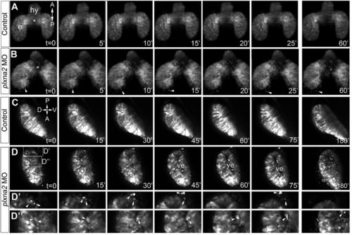
Retinal progenitors leave the eye vesicle with Plxna2 knockdown. Frames from time-lapse confocal imaging of control (A,C) and plxna2 morphant (B,D) rx3:GFP eyes viewed dorsally between 10 and 12 somites (A,B) and laterally between 8 and 14 somites (C,D). In the plxna2 morphant the eye field separates, but some cells leave the eye vesicle (arrowhead), whereas others fail to enter the eye vesicle (*). (D2,D3) Higher magnification view of the areas boxed in D, showing eye cells moving out of the eye (D2, arrows and arrowheads) or into the eye vesicle ventricle (ve) (D3, arrowheads). hy, hypothalamus. |
Expression Data
| Gene: | |
|---|---|
| Fish: | |
| Knockdown Reagent: | |
| Anatomical Term: | |
| Stage Range: | 5-9 somites to 14-19 somites |
Expression Detail
Antibody Labeling
Phenotype Data
| Fish: | |
|---|---|
| Knockdown Reagent: | |
| Observed In: | |
| Stage Range: | 5-9 somites to 14-19 somites |
Phenotype Detail
Acknowledgments
This image is the copyrighted work of the attributed author or publisher, and
ZFIN has permission only to display this image to its users.
Additional permissions should be obtained from the applicable author or publisher of the image.
Full text @ Development