
Fig. 2
- ID
- ZDB-FIG-140626-32
- Publication
- West et al., 2014 - Unusual Fluorescent Granulomas and Myonecrosis in Danio Rerio Infected by the Microsporidian Pathogen Pseudoloma Neurophilia
- Other Figures
- All Figure Page
- Back to All Figure Page
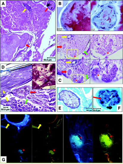
Histopathologic analysis of microsporidia-infected D. rerio. Zebrafish with fluorescent nodules and other signs of infection were analyzed with a panel of histologic stains: (A) H&E stain of fish cross section. Two granulomatous lesions surrounded by inflammatory cells are marked with red arrows. Lesion at upper right contains melanin pigment; yellow arrow denotes a blood vessel. (B) Luna stains of two granulomas. Red spherical objects within lesions are chitin-containing spores stained by Luna. Proteinaceous material stained red by Luna in granulomas is degenerating skeletal muscle. (C) H&E (top) and PAS (bottom) stains of spinal cord demonstrating classic Pseudoloma neurophilia lesions. Low-power images are at left; high-power images of yellow-boxed regions are at right. Spores in lesions appear as punctate basophilic spheres. Red arrows highlight a second lesion outside boxed regions; red asterisks denote two lesions in boxed areas. Yellow and green arrows denote spinal canal and meninges, respectively. (D) Trichrome stain of microsporidial lesions. Skeletal muscle stains pink (yellow arrowhead: longitudinal fibers; yellow arrow: cross-sectioned fibers) and connective tissue is blue. Red arrow identifies two granulomas with dark pink degenerating muscle and blue connective tissue remnants (green arrow). Inset panel displays spores on high power (1000×). (E) PAS stain of a granuloma surrounded by skeletal muscle, counterstained green. (F) GMS stain of a granuloma, highlighting carbohydrates in microsporidial cell walls. Lesion is GMS (+), but spores (red arrow) are GMS (). (G) Fungi-Fluor stain of fish cross section at low (left panels) and high power (right panels). Lesion (red asterisk) is blue with DAPI filter or yellow-green due to Fungi-Fluor staining of chitin in microsporidia with TRITC filter. Yellow arrow denotes a vertebral body adjacent to the peritoneal cavity. DAPI, 4,6-diamidino-2-phenylindole; H&E, hematoxylin and eosin; GMS, Grocott′s methenamine silver; PAS, Periodic acid Schiff; TRITC, tetramethylrhoodamine-5-isothiocyanate. |