
Fig. 5
- ID
- ZDB-FIG-140130-36
- Publication
- Fleming et al., 2013 - Functional characterisation of the maturation of the blood-brain barrier in larval zebrafish
- Other Figures
- All Figure Page
- Back to All Figure Page
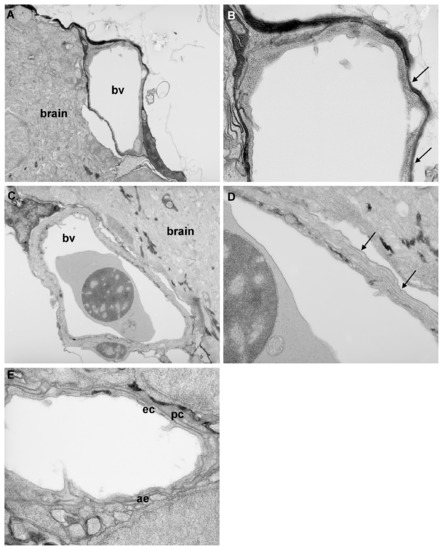
Transmission electron microscopy analysis of blood-brain barrier maturation. Parasaggital sections through the brain of zebrafish larvae at 3 d.p.f. and 10 d.p.f. A) At 3 d.p.f., blood vessels (bv) surrounding the brain are simple in structure. B) At high resolution, only a single membrane is observed (arrows) no evidence of double membranes was observed at any location examined. C) At 10 d.p.f., blood vessels surrounding the CNS are more complex in structure. D) At high resolution, a double layer membrane is apparent (arrows), indicative of the presence of tight junctions. E) In some vessels at 10 d.p.f., pericytes (pc) could be observed surrounding endothelial cells (ec). In addition, astrocyte endfeet (ae) were observed around some vessels at 10 d.p.f. but not at earlier ages. Magnification: A & C 5.2K; B, D & E 15.5K. |