
Fig. 2
- ID
- ZDB-FIG-131220-28
- Publication
- Yasuda et al., 2013 - A cis-acting element in the coding region of cyclin B1 mRNA couples subcellular localization to translational timing
- Other Figures
- All Figure Page
- Back to All Figure Page
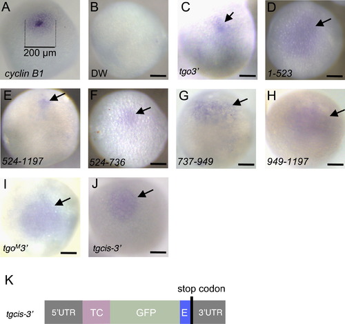
Localization of cyclin B1 reporter mRNAs that were transcribed from reporter genes injected into oocyte nuclei. (A) Whole-mount in situ hybridization of full-grown oocytes probed with cyclin B1. Aggregation of cyclin B1 mRNAs is found in the region indicated by dotted lines (200 μm). ((B)–(J)) Whole-mount in situ hybridization probed with gfp. Shown are oocytes injected with distilled water (DW) (B), reporter genes containing the full length (tgo3′) (C), 1–523 (D), 524–1197 (E), 524–736 (F), 737–949 (G) and 949–1197 (H) nts of the cyclin B1 coding region, tgoM3′ reporter gene (I) and tgcis-32 reporter gene (J). The oocytes injected with distilled water showed no signals (B). The tgo3′, 524–1197, 524–736 and tgcis–3′ mRNAs were aggregated in the animal polar cytoplasm ((C), (E), (F), (J)). In contrast, the 1–523, 737–949, 949–1197 and tgoM32 mRNAs were dispersed in the animal hemisphere of oocytes ((D), (G), (H), (I)). Arrows indicate the signals of reporter mRNAs. Bars, 100 µm. (K) Structure of the tgcis-3′ reporter gene, which contains cyclin B1 5′ UTR (52UTR), coding sequences of TC-tag (TC) and EGFP (GFP), CAGGAGACC element (E), a stop codon and cyclin B1 3′ UTR (3′UTR). |
Reprinted from Developmental Biology, 382(2), Yasuda, K., Kotani, T., and Yamashita, M., A cis-acting element in the coding region of cyclin B1 mRNA couples subcellular localization to translational timing, 517-29, Copyright (2013) with permission from Elsevier. Full text @ Dev. Biol.