
Fig. S4
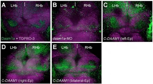
Changes in the relative levels of the endogenous Daam1a protein after local electroporation (Ep). (A) The Hb of control embryos labelled through indirect immunofluorescence against Daam1a (green) and counter stained with TO-PRO-3 to delineate the cellular context of the Hb (purple) showed a distinct punctate expression of Daam1a in the habenular neuropil, larger on the left compared to the right side at 96 hpf. (B) The Hb of embryos subjected to left-sided local electroporation of daam1a-MO show decreased levels of Daam1a expression at 96 hpf, primarily on the left Hb. (C) The Hb of embryos subjected to left-sided local electroporation of C-DAAM1 showed increased levels of Daam1a expression in the left Hb at 96 hpf. (D) The Hb of embryos after right-sided local electroporation of C-DAAM1 showed increase expression levels of Daam1a in the right Hb at 96 hpf, compared to controls. (E) The Hb of embryos subjected to bilateral local electroporation of C-DAAM1 showed increased levels of Daam1a expression in both left and right Hb at 96 hpf. Images correspond to dorsal views of maximum intensity z-stack confocal projections, with anterior to the top. Scale bar, 20 μm. |