
FIGURE
Fig. S2
- ID
- ZDB-FIG-131105-16
- Publication
- Satou et al., 2013 - Transgenic tools to characterize neuronal properties of discrete populations of zebrafish neurons
- Other Figures
- All Figure Page
- Back to All Figure Page
Fig. S2
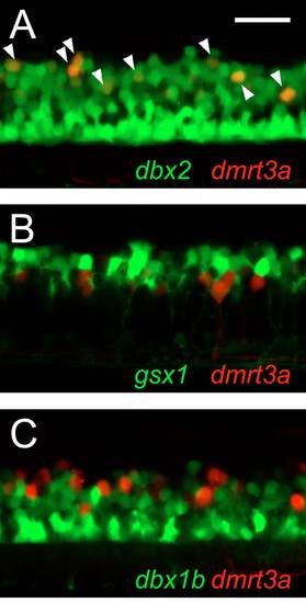
dmrt3a-positive neurons derive from the pd6 domain. Images are lateral views of the spinal cord at 3.5 dpf. (A)Tg[dbx2:GFP] and Tg[dmrt3a:RFP] compound transgenic fish. All the dmrt3a:RFP neurons are positive for dbx2:GFP (arrowheads). (B) Tg[gsx1:GFP] and Tg[dmrt3a:RFP] compound transgenic fish. None of the dmrt3a:RFP neurons is positive for gsx1:GFP. (C) Tg[dbx1b:GFP] and Tg[dmrt3a:RFP] compound transgenic fish. None of the dmrt3a:RFP neurons is positive for dbx1b:GFP. Scale bar: 50 μm. |
Expression Data
Expression Detail
Antibody Labeling
Phenotype Data
Phenotype Detail
Acknowledgments
This image is the copyrighted work of the attributed author or publisher, and
ZFIN has permission only to display this image to its users.
Additional permissions should be obtained from the applicable author or publisher of the image.
Full text @ Development