Fig. 4
- ID
- ZDB-FIG-130909-5
- Publication
- Heinke et al., 2013 - Antagonism and synergy between extracellular BMP modulators Tsg and BMPER to balance blood vessel formation
- Other Figures
- All Figure Page
- Back to All Figure Page
|
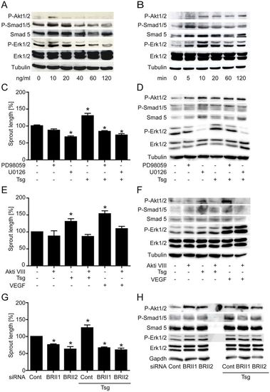
Recombinant Tsg protein induces concentration- and time-dependent intracellular signalling necessary for endothelial cell sprouting. (A,B) Dose and time dependence of intracellular signalling by Tsg in HUVECs. Western blot analyses were performed with the indicated antibodies. (C,E) Cumulative sprout length of HUVEC capillary-like structures from tube formation assays. (C) Tsg-stimulated sprouting was inhibited when Erk was blocked with signalling pathway inhibitors PD98059 (20μM) and U0126 (10μM) or (E) when Akt signalling was blocked with Akti VIII (2μM). Values are means ± s.e.m.; n = 3; *P<0.01 versus control. (D,F) Western blot analysis of Tsg-stimulated HUVECs with Erk inhibitors PD98059 (20μM) and U0126 (10μM) or Akt inhibitor Akti VIII (2μM) were performed with the indicated antibodies to determine the effects of inhibition of (D) Erk or (F) Akt phosphorylation on the other signalling pathways. (G,H) HUVECs were transfected with either of two BMPRII-specific siRNAs or scrambled siRNA control. (G) A tube formation assay was performed 48hours post-transfection and cumulative sprout length was measured. Values are means ± s.e.m.; n = 4; *P<0.01 versus siRNA control. (H) 48hours post-transfection cells were lysed and subjected to western blot analysis performed with the indicated antibodies. Representative western blots of one of three independent experiments are shown. |

