
Fig. S2
- ID
- ZDB-FIG-130907-35
- Publication
- McCarthy et al., 2013 - Pdgfra protects against ethanol-induced craniofacial defects in a zebrafish model of FASD
- Other Figures
- All Figure Page
- Back to All Figure Page
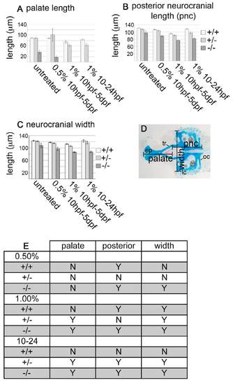
Ethanol-treatment causes a decrease in overall neurocranial length and width in pdgfra mutants. (A-C) Graphs depict the average (A) palate length, (B) posterior neurocranial length and (C) neurocranial width in μm. x-axis on all graphs indicate either untreated, 0.5% ethanol at 10 hpf-5 dpf, 1% ethanol from 10 hpf to 5 dpf, or 1% ethanol from 10 hpf to 24 hpf [standard error bars are depicted at 1.5 s.e.m., non-overlapping bars indicate significant differences (Moses, 1987)]. Wild type, light bar; pdgfra heterozygote, light-gray bar; mutant, dark-gray bar. (D) Alcian- and Alizarin-stained 5 dpf untreated wild-type neurocranium. Palate length was measured from the most anterior part of the ethmoid plate (ep) to the ends of the trabeculae (tr). The posterior neurocranium was measured from the most posterior part of the occipital arches (oc) to the ends of the trabeculae (tr). Neurocranial width was measured between the most lateral edges of the lateral commissures (lc). (E) Statistical analyses comparing the genotype/ethanol interactions with individual genotype and treatment controls. ‘Y’ indicates a significant difference. Wild-type (+/+) comparisons indicate whether there is a difference between treated and untreated wild-type embryos. pnc, posterior neurocranium. |
| Fish: | |
|---|---|
| Condition: | |
| Observed In: | |
| Stage: | Day 5 |