FIGURE

Fig. S1

ID
ZDB-FIG-130830-14
Publication
Dray et al., 2013 - Cell-Fibronectin Interactions Propel Vertebrate Trunk Elongation via Tissue Mechanics
Other Figures
All Figure Page
Back to All Figure Page
Fig. S1

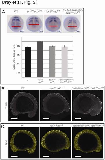
Analyses of Convergence Extension, Apoptosis, and Cell Proliferation in itgα5mo;αVmo Embryos, Related to Figure 1

(A) Wild-type, fn1mo;fn1bmo, itgα5mo;αVmo and Tg(tbx6l:itgα5-RFP);itgα5mo;αVmo embryos were fixed at the tailbud stage and in situ hybridization for her1 was performed. We then measured the medial-lateral width of the tailbud at the level of the posterior tip of the notochord. The fn1mo;fn1bmo embryos (n=30) exhibited a 17.5% increase in width of the tailbud (unpaired t-test; p-value <0.05) which is consistent with previous observations [1]. In contrast, neither itgα5mo;αVmo (n=37) nor Tg(tbx6l:itgα5-RFP);itgα5mo;αVmo embryos (n=24) displayed a change in tailbud width relative to wild type (n=55) (p-value >0.05). Error bars show standard error. The fn1 and fn1b morpholinos have been previously described [2].
(B) Cell death was assayed in wild-type, itgα5mo;αVmo and Tg(tbx6l:itgα5-RFP);itgα5mo;αVmo embryos. We fixed embryos at the 13-somite stage and assayed apoptosis via TUNEL. Briefly, embryos were fixed overnight in 4% PFA in PBS at 4°C and stored in 100% methanol at 20°C. Embryos were rehydrated and permeabilized in 100% acetone at 20°C for 20 min followed by several washes in 0.5% Triton X-100 and 0.1% sodium citrate in PBS and then treated for TUNEL assay using the in situ Cell Death Detection Kit, POD (Roche, Basel, Switzerland) according to the manufacture’s instructions. There was no clear difference in apoptosis in itgα5mo;αVmo embryos (n=12) nor in Tg(tbx6l:itgα5-RFP);itgα5mo;αVmo embryos (n=10) compared to wild type (n=12).
(C) Cell proliferation was assayed in wild-type, itgα5mo;αVmo and Tg(tbx6l:itgα5-RFP);itgα5mo;αVmo embryos. We fixed embryos at the 13-somites stage and assayed for cell proliferation via immunohistochemistry for Phosphorylated Histone H3 (PHH3) as previously described [3]. Compared to wild-type (n=5), itgα5mo;αVmo embryos (n=5) exhibited a 36% decrease in cell proliferation (unpaired t-test, p-value <0.05). Proliferation in Tg(tbx6l:itgα5-RFP);itgα5mo;αVmo embryos (n=5) was no different than in itgα5mo;αVmo embryos (p>0.05). Scale bars equal 200 microns.

Expression Data

Expression Detail
Antibody Labeling
Phenotype Data

Phenotype Detail
Acknowledgments
This image is the copyrighted work of the attributed author or publisher, and ZFIN has permission only to display this image to its users. Additional permissions should be obtained from the applicable author or publisher of the image. Full text @ Curr. Biol.