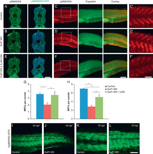
Knockdown of sulf1 in embryonic zebrafish leads to an increase in BMP signaling that can be rescued with inhibition of BMPRI. (A and B) Cross-sections through 36 hpf control (A) or sulf1 morphant (B) zebrafish labeled with anti-pSmad1/5/8 (green) and counterstained with DAPI. sulf1 morphants show stronger immunoreactivity against pSmad1/5/8 particularly around the spinal cord (arrow) and in the ventral regions of the somitic muscle near the notochord (brackets). (C and D) Whole-mount control (C), or sulf1 morphant (D) zebrafish fixed at 24 hpf, and double-labeled for pSmad1/5/8 and engrailed, with images taken at the level around and just posterior to the proctodeum. Control fish show discrete labeling of pSmad reactive nuclei in each somite, but absent from the horizontal myoseptum, where the engrailed-positive muscle pioneers can be found. sulf1 morphants show an increase in the level of pSmad immunoreactivity, and less organization of the pSmad immunoreactive cells particularly in the ventral portion of the more posterior somites, and with less separation between the dorsal and ventral halves of the domains of expression. Again, the number and intensity of engrailed-positive muscle pioneers is reduced in sulf1 morphants. (C′–D′) Magnified view of pSmad immunoreactivity from the boxed regions indicated in C and D. (E and F) Treatment of sulf1 morphant fish with 2 µM LDN193189 beginning at 12 hpf abrogates the increase in pSmad labeling, in cross-sections (E) and whole-mounts (F; magnified in F′). Treatment of sulf1 morphants with 2 μM LDN193189 is sufficient to reduce the levels of pSmad immunoreactivity, particularly in the ventral domain, consistent with inhibition of BMP signaling, and increase the number and intensity of muscle pioneers and medial fast cells. (G and H) Quantification of the number of muscle pioneer cells (G) and medial fast fibers (H) per somite in control (blue), sulf1 morphants treated with DMSO as a vehicle control (green), and sulf1 morphants treated with 2 μM LDN193189 beginning at 12 hpf. LDN treatment restored the number of MPCs to normal and led to a significant increase in the number of MFFs compared to DMSO treated morphants. (I–L) GFP expression under control of repeated BMP response elements in control (I and K) or sulf1 morphant (J and L) at 24 hpf (I and K) or 72 hpf (K and L). While controls have a distinct absence of GFP along the developing horizontal myoseptum, morphants show a greatly reduced gap between the dorsal and ventral expression, with some somites showing strong BMP activity along the lateral midline. Scale bars: A, B, E: 50 μm; C, D, F: 100 μm; C′, D′, F′: 50 μm; I–L: 100 μm.
|
