
FIGURE
Fig. 5
- ID
- ZDB-FIG-130510-34
- Publication
- Nevis et al., 2013 - Tbx1 is required for second heart field proliferation in zebrafish
- Other Figures
- All Figure Page
- Back to All Figure Page
Fig. 5
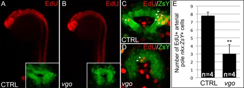
SHF progenitor cells fail to proliferate in the absence of Tbx1. A–D: Click-iT EdU labeling in 23ss Tg(nkx2.5:ZsYellow) vgo and control siblings. A,B: Flourescence microscopy images of EdU+ cells (red) in control (A) and vgo (B) embryos. 10× magnification, anterior up, dorsal right. Insets show flattened confocal images of ZsY+ cells comprising the cardiac cone. C,D: Composites of two confocal sections showing EdU+ cells (red) within the ZsYellow+ (green) SHF (white arrowheads). E: Graph depicting the average total numbers of EdU+, ZsYellow+ cells in confocal stacks of control (n=4) and vgo (n=4) embryos. |
Expression Data
| Gene: | |
|---|---|
| Fish: | |
| Anatomical Term: | |
| Stage: | 20-25 somites |
Expression Detail
Antibody Labeling
Phenotype Data
| Fish: | |
|---|---|
| Observed In: | |
| Stage: | 20-25 somites |
Phenotype Detail
Acknowledgments
This image is the copyrighted work of the attributed author or publisher, and
ZFIN has permission only to display this image to its users.
Additional permissions should be obtained from the applicable author or publisher of the image.
Full text @ Dev. Dyn.