FIGURE

Fig. S3

ID
ZDB-FIG-130502-13
Publication
Song et al., 2013 - Pou5f1-dependent EGF expression controls e-cadherin endocytosis, cell adhesion, and zebrafish epiboly movements
Other Figures
All Figure Page
Back to All Figure Page
Fig. S3

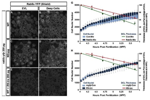
Comparison of Plasma Membrane Distribution of Rab5c-YFP and Number of Cell Nuclei during Early Epiboly
(Related to Figure 3)
(A-F) Confocal anti-GFP immunofluorescence images of Rab5c-YFP expressed by mRNA injection in WT (A-B) or MZspg embryos (C-D), and of Rab5c-YFP and RN-tre co-expressed in WT (E-F) embryos by mRNA co-injection. Animal views. Scale bar = 10 μm.
(G-H) Quantification of cell nuclei number and thickness of deep cell layer (DCL) from two hour (sphere to 50% epiboly) 3D time-lapse recordings of standard control morpholino (Con Mo) injected (n= 6 embryos), mgfp mRNA injected (n= 7 embryos), Rab5c morpholino (Rab5c Mo) injected (n= 7 embryos), or RN-tre mRNA injected (n= 5 embryos) WT embryos in which all cell nuclei were labeled by expression of NLS-tomato. Each embryo is documented by time series of animal pole view confocal stacks 120 μm from the animal pole EVL into the blastoderm. Cell nuclei numbers from the 3D time-lapse volume data in Rab5c Mo injected embryos (G) or RN-tre mRNA injected embryos (H) are not significantly different from control WT embryos, while thickness of the DCLs at 5.5 hpf as percentage 8 of DCL thickness at 4 hpf are significantly thicker both in Rab5c Mo (p < 0.05) and RN-tre mRNA (p < 0.01) injected embryos.
Boxplots and trend lines represent cell nuclei number and DCL thickness, respectively. Error bars represent standard error of the mean (SEM).

Expression Data

Expression Detail
Antibody Labeling
Phenotype Data

Phenotype Detail
Acknowledgments
This image is the copyrighted work of the attributed author or publisher, and ZFIN has permission only to display this image to its users. Additional permissions should be obtained from the applicable author or publisher of the image.

Reprinted from Developmental Cell, 24(5), Song, S., Eckerle, S., Onichtchouk, D., Marrs, J.A., Nitschke, R., and Driever, W., Pou5f1-dependent EGF expression controls e-cadherin endocytosis, cell adhesion, and zebrafish epiboly movements, 486-501, Copyright (2013) with permission from Elsevier. Full text @ Dev. Cell