
Fig. 4
- ID
- ZDB-FIG-130403-4
- Publication
- Tan et al., 2013 - Spatiotemporal expression of the dermatopontin gene in zebrafish Danio rerio
- Other Figures
- All Figure Page
- Back to All Figure Page
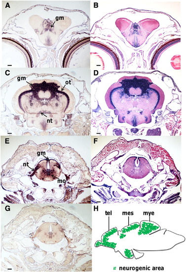
In situ hybridization of dermatopontin (DPT) mRNA in zebrafish brain. (A, C, E) In situ hybridization with DPT antisense probe. Transverse sections through the telencephalon, mesencephalon, and myelencephalon, respectively. (B, D, and F) Hematoxylin and eosin staining in the brain, counterpart to A, C, and E. (G) In situ hybridization with the DPT sense probe in the myelencephalon. (H) Schematic drawing of the neurogenic zone in the adult zebrafish brain (green areas). Abbreviations: gm, gray matter; ot, optic tectum; nt, neural tract; mo, medulla oblongata; tel, telencephalon; mes, mesencephalon; mye, myelencephalon. Scale bar = 100 μm. |
| Gene: | |
|---|---|
| Fish: | |
| Anatomical Terms: | |
| Stage: | Adult |
Reprinted from Gene, 516(2), Tan, Y., Iimura, K., Sato, T., Ura, K., and Takagi, Y., Spatiotemporal expression of the dermatopontin gene in zebrafish Danio rerio, 277-284, Copyright (2013) with permission from Elsevier. Full text @ Gene