
Fig. 4
- ID
- ZDB-FIG-130326-31
- Publication
- Ravi et al., 2013 - Sequencing of pax6 Loci from the elephant shark reveals a family of pax6 genes in vertebrate genomes, forged by ancient duplications and divergences
- Other Figures
- All Figure Page
- Back to All Figure Page
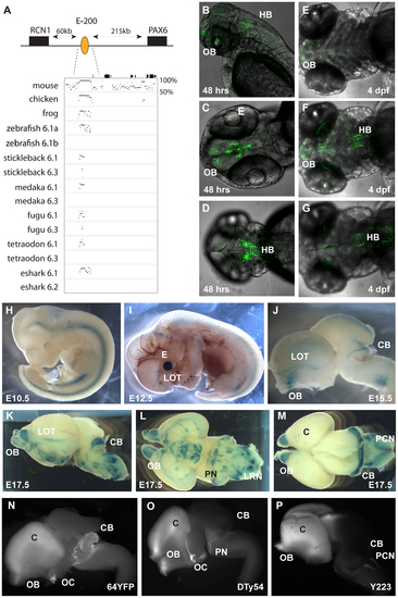
Characterization of the E-200 CNE in mouse and zebrafish reporter transgenics. A) The E-200 CNE is a deeply conserved cis-element found upstream of all vertebrate Pax6.1 loci and located about 200 kb upstream of human Pax6. A shorter stretch of the element is also conserved in the Pax6.3 loci of fugu and stickleback. (B–G) Fluorescent reporter transgenic zebrafish with the human E-200 element. B) Lateral view of an E-200-gata2-GFP reporter line showing expression in the olfactory bulbs (OB) and hindbrain (HB). C) Confocal image of the ventral head region of a 48 hpf transgenic fish reveals expression in the olfactory bulbs and the proliferative zones of the ventral brain region. D) Confocal view of the dorsal embryo head showing expression in the hindbrain. (E–G) Ventral, medial and dorsal confocal views show GFP signal in the olfactory bulbs, lateral olfactory tracts, forebrain (E, F) and hindbrain (F, G) of a 4 dpf reporter transgenic fish. (H–P) LacZ reporter transgenic mice with the human E-200 element. (H) At E10.5 no enhancer specific expression is observed. Signal in the neural tube is intrinsic to the hsp68-LacZ cassette, and in the genital ridge is aspecific. I) From E12.5 expression starts to appear in the olfactory tracts at the base of the cortex. J) At E15.5 staining is seen in the olfactory bulbs (OB), the lateral olfactory tracts (LOT) and a thin band in the dorsal cerebellum (CB). Expression is also seen in the precerebellar neuro-epithelium (PCN), migratory streams and the lateral reticular nuclei (LRN) of the hindbrain. K) This pattern is maintained at E17.5, with increased cerebellar expression covering the full width of the organ. L) Ventral view of an E17.5 dissected brain shows expression in the lateral olfactory tracts and in the precerebellar nuclei including the pontine gray nuclei (PN). Staining in hypothalamic areas was not seen in other transgenic lines, while expression in the olfactory bulbs varied in strength between lines. M) Dorsal view of an E17.5 brain shows the expression in the cerebellum and precerebellar neuroepithelium. N–P) Comparison of reporter YAC transgenic mice carrying 420 kb of the human PAX6 locus, which does not extend to include the E-200 long-range enhancer, to a targeted reporter insertion into the endogenous Pax6 locus shows a deficiency of reporter expression in the cerebellum of the YAC reporter lines. N) Fluorescence of a YFP reporter integrated into the endogenous mouse Pax6 gene is found in the olfactory bulbs (OB), cortex (C), optic chiasm (OC), cerebellum (CB) and pontine migratory stream (PMS). While reporter fluorescence signal of comparable strength is seen in both (O) single copy or multi-copy (N) YAC transgenic lines carrying 420 kb of the human PAX6 locus in the olfactory bulbs, cortical lobes, optic chiasma and pontine migratory streams, expression is absent from the cerebella of these transgenics. |
| Gene: | |
|---|---|
| Fish: | |
| Anatomical Terms: | |
| Stage Range: | Long-pec to Day 4 |