
Fig. 4
- ID
- ZDB-FIG-130313-6
- Publication
- Mongera et al., 2013 - Genetic lineage labeling in zebrafish uncovers novel neural crest contributions to the head, including gill pillar cells
- Other Figures
- All Figure Page
- Back to All Figure Page
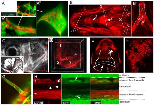
Detection of contested cranial NC derivatives. (A-A′′) MIP of confocal stacks showing labeled cartilaginous and bony elements in the otic capsule: chondroblasts and osteoblasts in the ventral cartilage (A′) and in the inner ear structures (A′′). (B) MIP of confocal stacks (ventral view) showing labeled viscerocranial structures: (1) Meckel′s cartilage (paired), (2) the basihyal (unpaired), (3) the ceratohyal (paired), (4) the basibranchial (unpaired) and (5) the branchiostegal rays (paired), with individual rays marked with an asterisk. (B′) Confocal section from B showing labeled (2) basihyal (unpaired), (3) ceratohyal (paired) and (4) basibranchial (unpaired) elements. (C,D) Epifluorescent image of mCherry+ osteoblasts forming the opercular series in Tg(sox10:ERT2-Cre;ubi:switch) adult fish (C) and Alizarin Red-stained skull (lateral view) in a 3-month-old zebrafish showing the four bones forming the opercular series: (1) preopercle, (2) interopercle, (3) subopercle and (4) opercle (inset). (D) 3D view of confocal stacks (boxed region in C), which shows positive cells in all the bones composing the opercular series. (E) Frontal (Fr) and parietal (P) bones. Epifluorescence image showing recombined osteoblasts in the rostral part of the frontals (arrowhead), whereas the parietals are devoid of labeled osteoblasts (arrow indicates a pigment cell clone). (F) MIP of confocal stacks showing labeled osteoblasts in the anterior region of the frontals. (G) MIP of confocal stacks of a maxillary barbel in induced adult fish showing recombined tissues in the core of the tactile organ. (H) Confocal section showing, in the red channel, labeled nerves (asterisks), fibroblast-like cells in the rod matrix (arrowheads) and flattened cells along the dorsal aspect of the rod (arrow). In the green channel, a non-switched lymphatic vessel (arrow) and blood vessel (arrowhead) are indicated. In the merge panel, the different layers composing the barbels are shown. |