
Fig. S3
- ID
- ZDB-FIG-130225-11
- Publication
- Dong et al., 2012 - Suppression of rap1 impairs cardiac myofibrils and conduction system in zebrafish
- Other Figures
- All Figure Page
- Back to All Figure Page
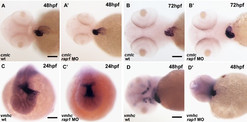
The cardiac looping defect was also evident by in situ hybridization. At 24 hpf, both control and rap1MO animals showed alike vmhc expression pattern in their hearts. Unlike the wild type hearts (A, C, D), the heart tube failed to extend left (C, C′) at 24 hpf or failed to form ‘S’ loop (A, A′, D and D′) in rap1MO animals at 48 hpf. At 72 hpf, ventricular defects, including pericardium edema and abnormal chamber differentiation, was observed in rap1MO (B, B′′). Ventral views were shown in A-A′, B-B′ and D-D′, anterior to the left;.dorsal view are shown in C-C′, anterior to the bottom. Scale bar, 150 μm in A, A′, D and D&prime& 120 μm in B and B&prime& 80 μm in C and C′. |