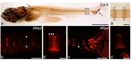
Early mineralization stages of vertebral centra and association with cartilaginous arches. Lateral view of (a, a′) Collagen type II immunostaining in the vertebral column of a 5.9 mm TL zebrafish shows (a) protein accumulation mostly in the cartilaginous structures of the Weberian apparatus and of the caudal fin skeleton (arrows) but also in the notochord sheath (white arrowheads). (a′) Although less evident than in cartilage, the staining can be clearly identified in the notochord matrix in a segmented manner (white arrowheads). (b-c) Early mineralization can be detected through alizarin red S (ARed) staining viewed under fluorescent light. (b) Early stage “ring” centrum (arrowheads) and (b′) close up showing the mineralization fronts (white arrowheads) in 5.0 mm TL fish. (c) Caudal fin centrum mineralization with a basiventral origin (arrowhead) and (c′) close up presenting a uniform mineralization surface with no distinct mineralization fronts, here represented by a 6.5 mm TL fish. Scale bars (a): 1 mm; (a′) 0.1 mm; (b,c): 0.15 mm; (b′, c′) 30 μm.
|
