
Fig. s1
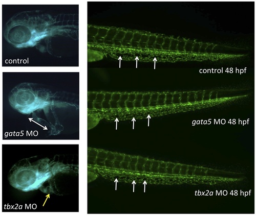
The CHT defect caused by depletion of Gata4 is not seen in other models of embryonic cardiomyopathy. The heart and caudal tail regions are shown in representative 48 hpf tg(gata1:dsred; fli1:egfp) uninjected control embryo (top), or embryos that had been injected with morpholinos targeting gata5 (middle) or tbx2a (bottom). In the trunk images, white arrows indicate examples of characteristic fenestrated structures that start to form around 32 hpf in the CHT plexus, but fail to do so in the gata4 morphant. The control embryo has a normally looped heart, while the gata5 morphant has an unlooped heart-string (indicated by the double-headed arrow), and the tbx2a morphant has an extended dysmorphic atrium (indicated by the yellow arrow). Morpholinos were used under conditions that generate reproducible cardiomyopathies and poor circulation with pericardial edema, as documented in our own and others studies: gata5, 52-AAGATAAAGCCAGGCTCGAATACAT, 10 ng/embryo; tbx2a,52-CGGTGCATCCAACAAACGTAGTGAA, 5 ng/embryo. |