
Fig. S8
- ID
- ZDB-FIG-130123-50
- Publication
- Schweitzer et al., 2013 - Sim1a and Arnt2 contribute to hypothalamo-spinal axon guidance by regulating Robo2 activity via a Robo3-dependent mechanism
- Other Figures
- All Figure Page
- Back to All Figure Page
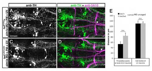
Medial displacement of TH-positive longitudinal axons in robo3 mutants is reduced after temporally controlled sim1a knockdown. Dorsal views of confocal z projections of the hindbrain of 72 hpf embryos labeled with anti-TH and anti-3A10 antibodies are shown. (A,B) Longitudinal TH-positive axons derived from group 2 and 4 DA neurons are shifted towards the midline (arrows in A) after photoactivation of sim1a MO at 22 hpf in twttw204/+ embryos. Midline crossing of Mauthner axons is normal (arrowhead in B). (C,D) After temporally controlled activation of sim1a MO in twttw204/tw204 embryos at 22 hpf, TH-positive longitudinal axons derived from group 2 and 4 DA neurons project in a wild-type-like fashion (arrows in C). Abnormal crossing of Mauthner axons (arrowhead in D) indicates homozygous robo3 mutant. (E) Quantification of medio-lateral positioning of TH-positive longitudinal axons in twttw204/+ and twttw204/tw204 embryos after temporally controlled activation of sim1a MO. Asterisks in A and C indicate noradrenergic locus coeruleus neurons. PM, photomorph; UV, ultra violet. Numbers in A and C indicate DA neuronal groups according to nomenclature of Rink and Wullimann (Rink and Wullimann, 2002). *P< 0.001, Student’s t-test; n.s., not significant. Scale bar: 50 μm for A-D. |