
Fig. 11
- ID
- ZDB-FIG-130118-6
- Publication
- López-Bellido et al., 2012 - Cocaine Modulates the Expression of Opioid Receptors and miR-let-7d in Zebrafish Embryos
- Other Figures
- All Figure Page
- Back to All Figure Page
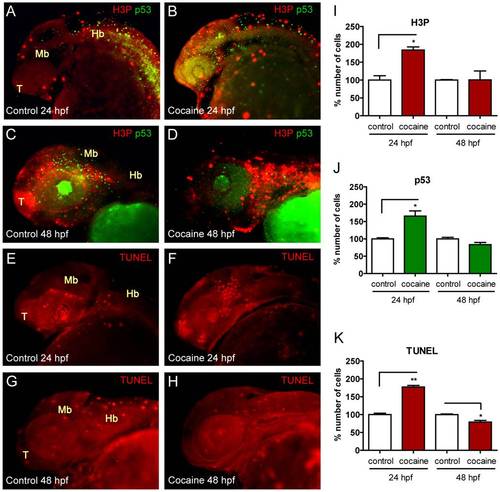
Analyses of proliferation and apoptosis in whole-mount embryos by IHC and TUNEL assays. Lateral views of embryos not exposed to cocaine are on the left side (A, C, E and G) and those exposed to the drug are on the right (B, D, F and H). Cocaine-exposed embryos showed an increase in apoptotic cells (p53) at 24 hpf (A vs. B) and a decrease at 48 hpf (C vs. D). This finding was similar with the TUNEL assay method at 24 hpf (E vs. F) and 48 hpf (G vs. H). The number of proliferating cells (in percent) (H3P) is increased both at 24 and 48 hpf (A vs. B and C vs. D, respectively). I, J and K show the percent changes in H3P, p53, and TUNEL at 24 and 48 hpf. Abbreviations: H3P: phosphorylated histone-3; p53: p53 protein; TUNEL: Terminal deoxynucleotidyl transferase dUTP nick end labeling; T: telencephalon; Mb: midbrain; Hb: hindbrain. Scale bars 150 μm. P-Values were calculated by two-tail unpaired student′s t test: *P<0.05, **P<0.01. |
| Fish: | |
|---|---|
| Condition: | |
| Observed In: | |
| Stage Range: | Prim-5 to Long-pec |