
Fig. 3
- ID
- ZDB-FIG-130108-17
- Publication
- Sharma et al., 2013 - PYK2: A calcium-sensitive protein tyrosine kinase activated in response to fertilization of the zebrafish oocyte
- Other Figures
- All Figure Page
- Back to All Figure Page
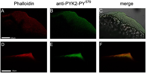
PYK2 activation co-localized with actin cytoskeletal reorganization. Oocytes were fixed at 1.0 (panels A–C) and 2.5 m.p.i.(panels D–F), then labeled with alexa 565—phalloidin (red) and anti-PYK2-PY579 followed by alexa 488-goat anti-rabbit IgG (green). Confocal images are displayed showing actin in the red channel (A and D), phosphorylated PYK2 in the green channel (B and E), or the merged red and green channels (C and F). Magnification is indicated by the bar which represents 100 µm. (For interpretation of the references to color in this figure legend, the reader is referred to the web version of this article.) |
| Antibody: | |
|---|---|
| Fish: | |
| Anatomical Term: | |
| Stage: | 1-cell |
Reprinted from Developmental Biology, 373(1), Sharma, D., and Kinsey, W.H., PYK2: A calcium-sensitive protein tyrosine kinase activated in response to fertilization of the zebrafish oocyte, 130-140, Copyright (2013) with permission from Elsevier. Full text @ Dev. Biol.